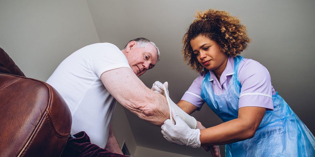
Skin tears are a common wound type and a growing problem, especially in New Zealand’s ageing population.
The limited research undertaken in New Zealand suggests there are inconsistencies in the use of skin tear wound management and prevention strategies in community and hospital settings.
As nurses, it is our responsibility to ensure we assess our clients/residents for the potential risk of skin tears, put in place strategies to minimise these risks, and if an injury does occur, then act promptly to assess and manage the skin tear appropriately. If the correct management of these skin tears is not undertaken, then there is the potential for these wound types to become chronic and non-healing.
An international consensus panel defined skin tears as: A wound caused by shear, friction, and/or blunt force resulting in separation of skin layers. A skin tear can be partial thickness (separation of the epidermis from the dermis) or full thickness (separation of both the epidermis and dermis from underlying structures). (LeBlanc and Baranoski, 2011)
Relevance to the New Zealand population
The overall prevalence of skin tears in New Zealand is unknown. However, ACC 2011 data reports that 63 per cent of ACC nursing services claims were classified as open wounds. More than 40 per cent of these were for people aged over 75 years and 8 per cent for people aged between 60 to 74 years.
In comparison, the Australian data shows that skin tears account for nearly 55 per cent of all wound types in the elderly and in the US, there is an estimated incidence level of 0.92 – 2.5 per patients per year accounting for approximately 1.5 million skin tears a year in adults in health care or aged care facilities.
These statistics suggest that the prevalence of skin tears in New Zealand is also high and with our ageing population we can expect the incident rate of this wound type to grow exponentially.
This claim is supported through research undertaken by research fellow Meg Butler, published in the New Zealand Medical Journal in 2004, where she looked at the prevalence of falls in New Zealand aged care facilities. From her data, it was shown that of the 954 falls reported over an 18-month period, a quarter (228) resulted in a skin tear.
Risk factors
Skin tears are associated with falls, blunt trauma, handling, and equipment injuries. A number of risk factors have been reported including:
- Dependence on others for activities of daily living (highest incidence)
- Hospital beds are the most common causes of traumatic-induced skin tear followed by the wheelchair (PA-PSRS Patient Safety, 2006)
- Intravenous catheters are the most likely of all drains and tubes to cause a skin tear (Baranoski, 2003)
- Radiography procedures were the highest risk procedure when transferring and positioning patients (PA-PSRS Patient Safety, 2006)
- Independent ambulatory patients (2nd highest incidence)
- Vision impaired patients (3rd highest incidence)
- Sensory changes/loss – e.g. hearing, sensation, vision
- Advanced age
- Immature skin (premature infants)
- Ageing females
- Multiple medications: steroids – systemic or topical, anticoagulants, polypharmacy
- History of previous skin tears
- Dry, fragile skin/ecchymoses (bruising/discolouration of the skin caused by leakage of blood into the subcutaneous tissue as a result of trauma to the underlying blood vessels)
- Poor nutrition and hydration
- Cognitive or sensory impairment/perception
- Co-morbidities that compromise vascularity and skin status, including chronic heart disease, renal failure, cerebral vascular accident, diabetes, immuno-compromised, hypoalbuminsm, hypothyroidism, or ureamia
- Impaired mobility/immobility/poor balance/poor locomotion
- Presence of friction, shearing or pressure
- Incorrect removal of adhesive dressings.
Location
Skin tears can occur on any part of the body. In the elderly, they are often sustained on the legs if mobile, arms if immobile, as well as the dorsal (back) of the hands.
Skin tears in neonates with immature skin tend to be associated with the use of adhesives or device trauma and often occur on the head, face and extremities
Diagnosis
The correct diagnosis and grading of a skin tear is vital to aid clinical management decisions.
To guide this assessment, the use of a validated assessment tool is recommended. In New Zealand and Australia, the STAR (Skin Tear Audit Research) classification system – developed by Professor Kerlyn Carville’s team in Western Australia – appears to be the system of choice. The use of a tool ensures that clinicians are using the same terminology to describe the degree of skin damage and this in turn will inform others to the correct degree of skin damage/loss (see classification sidebar).
Points to remember:
- Leave a space between each steri-strip to allow exudate to drain and to accommodate for swelling as part of the normal inflammatory response.
- Gently lay the steri-strips onto the periwound and then over onto the fragile flap; do not stretch the steri-strip, this could cause additional trauma and potentially flap ischemia.
- The secondary dressing needs to be non-adherent, absorbent and protective.
- Take care when applying adhesive tapes to fragile skin; there is the potential to cause additional trauma.
Don’t forget the tetanus shot: the human tetanus immunoglobulin should be given before wound debridement because exotoxin may be released during wound manipulation.
Predicting risk and prevention strategies
It is critical to predict and identify those at high risk for skin tears so that an appropriate prevention programme can be implemented before injury occurs.
In the New Zealand setting, there appears to be a lack of a validated tool to predict skin tear risk, and skin tears are not generally assessed using a validated assessment system. A team approach to assessment and implementation is vital to minimise the potential risk of skin tears.
Strategies in preventing skin tears
- Assess for risk upon admission to healthcare service and whenever the individual’s condition changes.
- Implement a systematic prevention protocol.
- Have individuals at risk wear long sleeves, long pants/trousers, or knee-high socks.
- Provide shin guards for those individuals who experience repeat skin tears to shins.
- Ensure safe patient handling techniques and equipment/environment.
- Involve individuals and families in preventive strategies.
- Educate registered and non-registered staff and caregivers to ensure proper techniques for providing care without causing skin tears.
- Consult dietitian to ensure adequate nutrition and hydration.
- Keep skin well lubricated by applying hypoallergenic moisturiser at least two times per day.
- Protect individuals at high risk from trauma during routine care and from self-injury.
Source: Le Blanc K and Baranoski S (2011), Skin tears: state of the science, consensus statements for the prevention, prediction, assessment and treatment of skin tears, Advances in Skin & Wound Care, 24(9)(Suppl 1):2-15
Conclusion
The very old and the very young (particularly nenonates with oxygen tubes or IV lures) are at the highest risk of skin tears. While skin tears may not be life-threatening, they are a painful injury that can also be disfiguring.
Preventing skin tears from happening in the first place should be our primary focus as nurses. We need to be knowledgeable about the risks and have the skills to assess and treat these wounds when they do occur. This requires being conscious that applying and removing adhesive tapes to fragile skin has the potential to cause additional trauma.
Consider not only using skin tear prevention strategies but also using a validated skin tear risk assessment tool. Such tools are commonly used in the United States and Australia.
Once a skin tear has occurred, using a standardised assessment classification system – such as STAR – to diagnose and grade a tear is a vital aid to managing the wound. Developing your own set of protocols for managing skin tears and ongoing staff education can help reduce the incidence and the time spent treating these too common wounds.
*Author: Wound consultant Elizabeth Milner is director of nursing for Geneva Healthcare and a wound care lecturer at the University of Auckland.
DRESSING SELECTION AND MANAGEMENT
Using a STAR acronym.
S STOP BLEEDING AND CLEAN
- Select cleansing product.
- Control bleeding (compression/elevation).
- Clean the wound bed.
T TISSUE ALIGNMENT
- If the skin flap is viable, roll skin flap back into place, using a moistened cotton bud.
- Secure the flap. Consider steri-strips to secure edges of flap(s).
- Leave a space between each strip to facilitate exudate drainage.
- DO NOT attempt to completely approximate edges.
- The flap should be left in place for approximately 5 days (unless dark or dusky – see below) to allow for the skin flap to “take”.
A ASSESS AND DRESS
- Assess the degree of tissue loss and skin flap colour using the STAR Classification system.
- Refer to general practitioner if surgical intervention required.
- Consider factors affecting wound healing.
- Inspect surrounding skin and environment for re-injury risk.
- Consider non-adhesive foam or silicone/adhesive foam products.
- Mark the outer dressing with an arrow towards the non-attached margin to indicate the direction in which to remove the dressing.
- Mark the date for removal.
- Apply a tubular bandage to facilitate haemostasis and for ongoing protection.
- Complete the Wound Chart and document interventions in progress notes.
R REVIEW AND RE-ASSESS
- If skin flap is dusky / darkened – reassess in 24 to 48 hours.
- Determine and document date of wound review in 5 days.
- Monitor for changes in wound status.
- Maintain skin integrity.
- Note: Unless wound assessment indicates otherwise, leave the primary dressing intact for 5 days or as per dressing recommendation. The secondary dressing may be changed as required.
References:
- Meg Butler, Ngaire Kerse, Maree Todd (2004), Circumstances and consequences of falls in residential care: the New Zealand story The New Zealand Medical Journal Vol 117 No 1202 ISSN 1175 8716
- Bank, D., & Nix, D. (2006). Preventing skin tears in a nursing and rehabilitation center: An interdisciplinary effort. Ostomy Wound Management, 52(9), 38-46.
- Carville, K., Newall, G., Haslehurt, P., Sanatmaria, R., & Roberts, N. (2007). STAR: A consensus for skin tear classification. Primary Intention, 15(1), 18-28.
- Le Blanc K and Baranoski S (2011), Skin tears: state of the science, consensus statements for the prevention, prediction, assessment and treatment of skin tears, Advances in Skin & Wound Care, 24(9)(Suppl 1):2-15
- LeBlanc, K., Christensen, D., Orsted, H., & Keast, D. (2008). Prevention and treatment of skin tears. Wound Care Canada, 6(1), 14-30.
- Baranoski S (2003) How to prevent and manage skin tears. Advances in Skin and Wound Care; 16: 5, 268-270.
- Bianchi J (2012) Preventing, assessing and managing skin tears. Nursing Times; 108: 13, 12-16.
- PA PSRS Patient Saf Advis 2006 Sep; 3(3):1,5-10.
